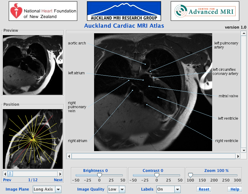
The Atlas images have been acquired in the standard image planes of axial, coronal and sagital to the cardiac specific planes of long and short axes, additionally images have been taken of the right ventricular outflow tract and of the aortic arch.
The AMRG Cardiac MRI Atlas is a complete labelled MRI image set of a normal patient's heart acquired with the Auckland MRI Research Group's Siemens Avanto scanner. The atlas aims to provide university and school students, MR technologists, clinicians and patients a tool for learning about the heart anatomy while becoming familiar with MRI, an important and growing tool in cardiology.
Data
There are 4 protocols used to create the cardiac atlas: T1-Weighted Images, True FISP Cines, MR tagging and contrast MRI. All acquired by using 1.5T SIEMENS Avanto MR machine. The T1-Weighted Images and True FISP Cines were acquired using a breath-hold retrospectively gated steady state free precession anatomical spin echo sequences with a phased array surface coil and ECG-R wave triggering. MR tagging requires the application of additional spatial modulating gradient, (cSPAMM) to create the image voids of the tags and the Phase Contrast MR's applies a velocity encoding gradient in the through plane direction.
| Age | 25 |
| Sex | Male |
| Height | 168 cm |
| Weight | 80.8 kg |
| Ethnicity | Indian |
| Smoker | No |
| Alcohol intake | No |
| Hypertension | No |
| Previous MI | No |
| Previous CABG | No |
| Previous PTCA | No |
| Angina | No |
| Heart Failure | No |
| Rheumatic Fever | No |
| Diabetes | No |
| Congenital Heart Disease | No |
| Renal Failure | No |
| Heart Rate | 72 beats/min |
| Systolic Blood Pressure | 106 mmHg |
| Diastolic Blood Pressure | 76 mmHg |
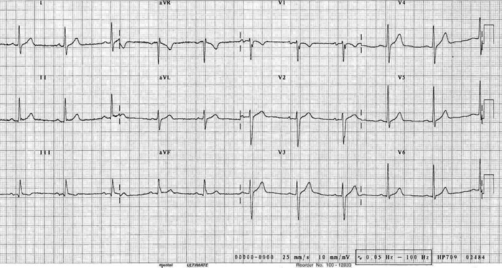
| Heart Rate | 62 beats/min |
| PR | 172 |
| QRSD | 98 |
| QT | 359 |
| QTc | 364 |
| T1 Weighted | True FISP Cine | Phase Contrast | Tagging | |
| Sequence | Spin Echo | Spin Echo | Gradient Echo | Gradient Echo |
| Slice thickness | 6 mm | 6 mm | 8 mm | 8 mm |
| TR | 944.1 ms | 27.2 ms | 28 ms | 37 ms |
| TE | 29 ms | 1.28 ms | 3.2 ms | 3.8 ms |
| Averages | 1 | 1 | 3 | 1 |
| Sampling | 100% | 100% | 80% | 75% |
| FOV | 100% | 81.25% | 75% | 81.25% |
| Num images | 1 | 32 | 25 | 20 |
| Flilp angle | 180 | 75 | 30 | 11 |
| Rows | 256 | 208 | 192 | 208 |
| Columns | 256 | 256 | 256 | 256 |
| Pixel Spacing | 1.37 x 1.37 mm | 1.25 x 1.25 mm | 1.37 x 1.37 mm | 1.33 x 1.33 mm |
Contributors
- Kieran O'Brien - Auckland Bioengineering Institute
- Alistair Young - Auckland MRI Research Group
- Brett Cowan - Centre for Advanced MRI
- Chris Occleshaw - Auckland City Hospital
- Sandra Winsor - Centre for Advanced MRI
- Shelley Park - Centre for Advanced MRI
- Gareth de Walters - Auckland Bioengineering Institute
If you would like to cite this data, use the following citation:
AMRG Cardiac Atlas, Auckland MRI Research Group, Auckland, New Zealand. Available online http://www.cardiacatlas.org/studies/amrg-cardiac-atlas/.